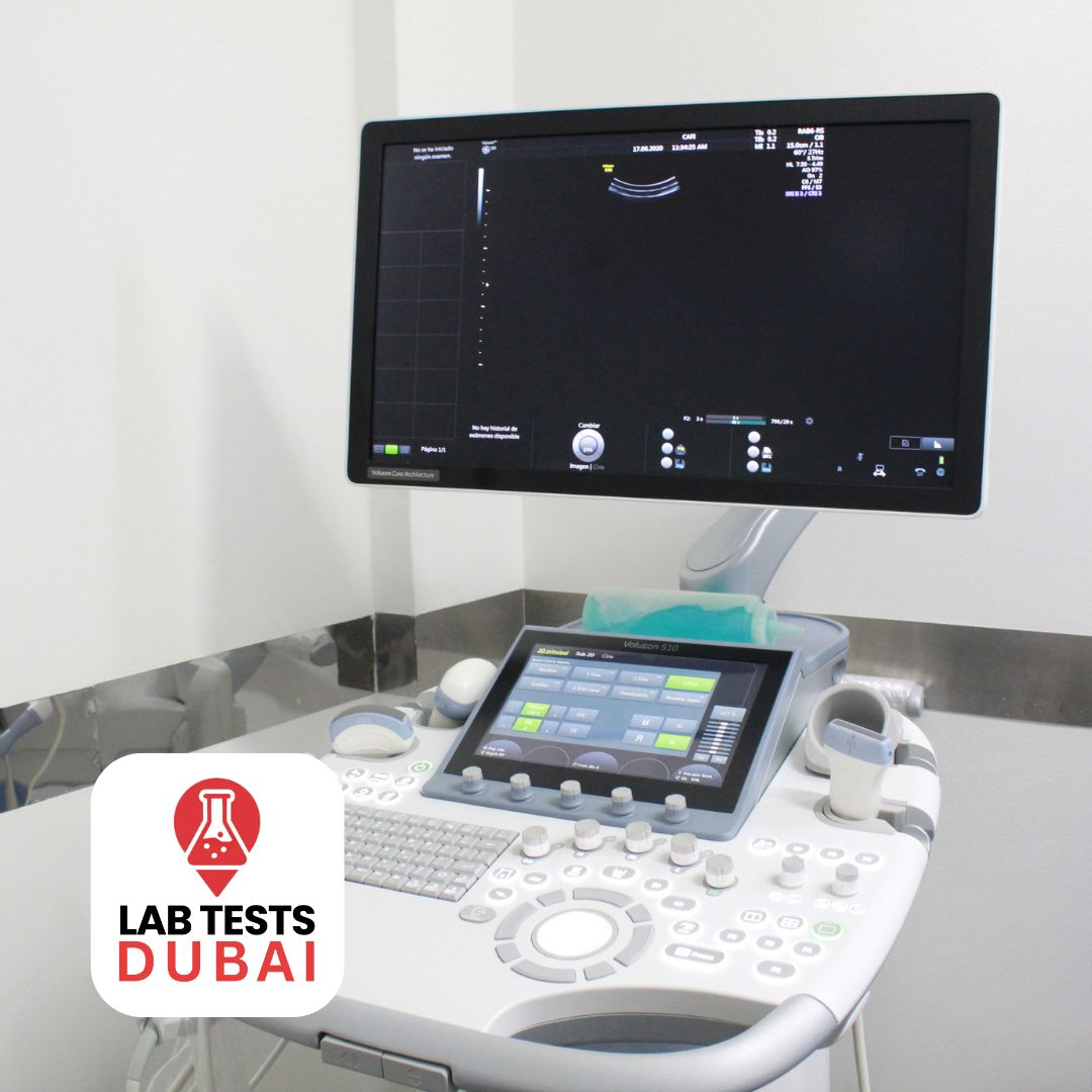
Abdo Back Wall Ultrasound in Dubai – Posterior Abdominal Imaging for Hernias & Lesions
550,00 د.إ
The US Exam Abdo Back Wall – Ultrasound is a specialized training resource aimed at improving ultrasound examination proficiency, particularly for evaluating the abdominal back wall. This tool offers realistic ultrasound imagery, high-quality anatomical features, and durable materials suitable for repeated practice. It enables medical professionals and students to refine their skills in probe positioning, image analysis, and diagnostic methods within a controlled setting. With its realistic echogenic qualities, it provides an authentic scanning experience, making it an invaluable asset for enhancing skills in abdominal ultrasound assessments. Perfect for medical training programs, it boosts confidence, accuracy, and diagnostic effectiveness in clinical settings.
Description
Abdo Back Wall Ultrasound in Dubai – Posterior Abdominal Imaging for Hernias & Lesions
The US Exam Abdo Back Wall – Ultrasound is a highly accurate imaging tool specifically designed for thorough evaluations of the abdomen. By employing cutting-edge ultrasound technology, this exam offers a clear view of the posterior abdominal wall, facilitating the identification of abnormalities, assessing structural integrity, and diagnosing pathological conditions.
Why You Need This Test
The US Exam Abdo Back Wall – Ultrasound is a specialized diagnostic imaging procedure that provides high-resolution, real-time visualization of the posterior abdominal wall—the deep muscular and fascial layer at the back of the abdomen.
This test is essential for:
- Detecting retroperitoneal or occult hernias (e.g., lumbar, obturator, or Spigelian hernias)
- Evaluating soft tissue masses, tumors, or abscesses
- Assessing muscle tears, hematomas, or post-surgical complications
- Investigating unexplained lower back or flank pain
- Monitoring abdominal wall integrity after surgery or trauma
Unlike standard abdominal ultrasounds, this exam focuses specifically on the back wall structures, offering critical insights that may be missed in routine scans.
Symptoms That Indicate This Test
You may need this ultrasound if you experience:
- Persistent pain in the lower back, flank, or side that worsens with movement
- A palpable lump or bulge in the lower abdominal or flank region
- Swelling or discomfort after abdominal surgery
- History of abdominal trauma or muscle strain
- Unexplained referred pain to the groin or leg
- Suspected rare or atypical hernia
This test is particularly valuable for athletes, post-surgical patients, and individuals with chronic back pain of unclear origin.
Natural Production & Imaging Principle
The posterior abdominal wall is composed of muscles (transversus abdominis, quadratus lumborum), fascia, and connective tissue that support internal organs and stabilize the trunk.
Ultrasound uses high-frequency sound waves—not radiation—to generate real-time images of soft tissues. With Doppler capabilities, it can also assess:
- Blood flow in surrounding vessels
- Inflammation or vascularity in masses
- Fluid collection or hematoma
Because it’s non-invasive and radiation-free, it can be used safely for repeated monitoring.
What Happens If Untreated
Ignoring abnormalities in the abdominal back wall can lead to:
- Progression of undiagnosed hernias, potentially leading to strangulation or bowel obstruction
- Chronic pain or muscle dysfunction
- Delayed diagnosis of tumors or infections
- Complications after surgery or trauma
Early detection ensures timely intervention—whether conservative management, physiotherapy, or surgical repair.
How to Prepare for the Test
Preparation is minimal:
- Wear loose, comfortable clothing
- No fasting required
- You may be asked to lie on your side or back during the scan
- Avoid applying lotions or oils to the lower back or flank area
The procedure takes 20–30 minutes and is completely painless.
Test Overview
| Parameter | Details |
| Test Name | US Exam Abdo Back Wall – Ultrasound |
| Imaging Type | Diagnostic Ultrasound (Sonography) with Doppler |
| Area Covered | Posterior Abdominal Wall (lumbar, flank, and lower abdominal regions) |
| Radiation Exposure | None – Completely radiation-free |
| Reporting | Certified radiologist-reviewed report with findings and recommendations |
Don’t let unexplained back or flank pain go unchecked. The Abdominal Back Wall Ultrasound gives you clear, precise imaging—helping detect hidden hernias, lesions, or muscle injuries.
👉 Book your US Exam Abdo Back Wall today with Lab Tests Dubai and receive professional imaging and results within 10 days.
See what’s beneath the surface. Diagnose with confidence. Heal with clarity.


Reviews
There are no reviews yet.